Question 1:
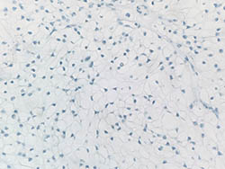
A 79-year-old healthy female with no prior history of cancer or skin disease presented to her dermatologist with a 0.7 x 0.5 x 0.5 cm exophytic red papule on her right neck. Clinical and histopathological examination of the biopsy revealed the following in figures 1-4. Immunohistochemical stains were performed including EMA, CEA, CD10, Vimentin, and RCC.
Click this image to view the virtual slide.
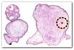
Figure 1.
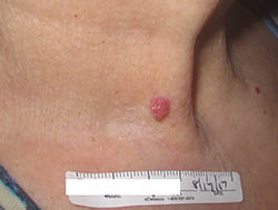
Figure 2.
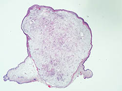
Figure 3.
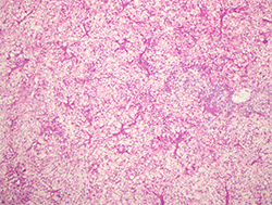
Figure 4.
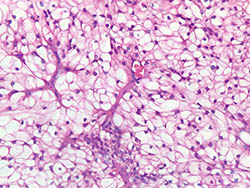
Figure 5. (EMA)
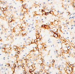
Figure 6. (Vimentin)
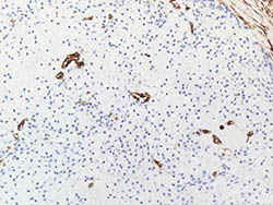
Figure 7. (CD10)
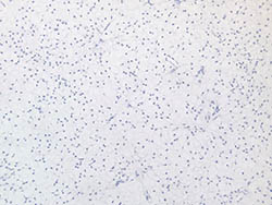
Figure 8. (RCC)
Which of the following is the best diagnosis?