Question 1:
An 80-year-old man presented with a 5-cm mass on his left upper lip that had been growing for more than 5 years. Pathologic examination of an excisional biopsy revealed the following:
Click this image to view the virtual slide
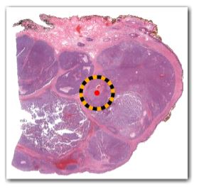
Figure I: Whole slide image of a representative section.
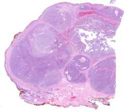
Figure II: H&E, 40x (A), 100x (B and C)
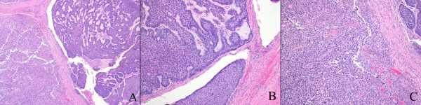
Figure III: H&E, 20x magnification
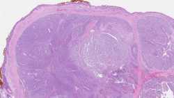
Figure IV: Immunohistochemical stains for cytokeratin-AE1/AE3 (A) and Melan-A (B), 40x magnification, same focus as Figure III
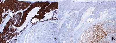
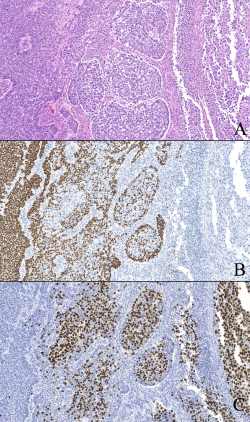
Figure V: H&E, 100x, central zone seen in Figure III
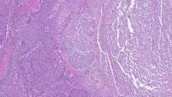
Figure VI: H&E (A) and immunohistochemical slides for p63 (B) and SOX10 (C), 200x magnification, same focus as Figure V
Which of the following is the best diagnosis?