Question 1:
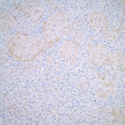
A 30-year-old man with a history of childhood leukemia presents with a sharply demarcated erythematous lesion on the back. The lesion has been present for five months. A biopsy is performed, showing the lesion below. Immunohistochemical staining reveals that the granular cells are negative for CD163 and S-100, but positive for CD68.
Click this image to view the virtual slide
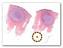
Figure 1. A low-power, overall view (12x).
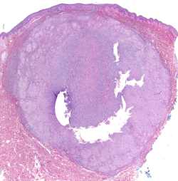
Figure 2. A low-power view (40x).
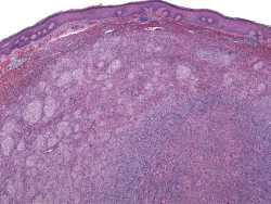
Figure 3. A medium-power view (100x).
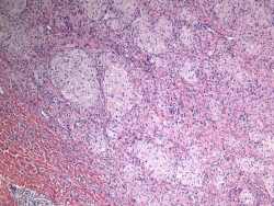
Figure 4. A high-power view (200x).
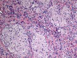
Figure 5. A high-power view of the edge of the lesion (200x).
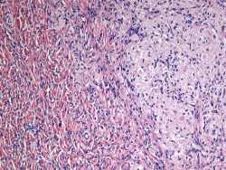
Figure 6. A higher-power view (400x).
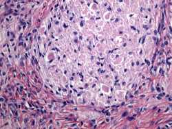
Figure 7. CD163 stain of the tumor (200x).

Figure 8. S-100 stain of the tumor (200x).
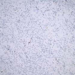
Figure 9. CD68 stain of the tumor (200x).
What is the most likely diagnosis?