Question 1:
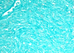
A 74-year-old man with a history of rheumatoid arthritis presents with a nodule on his wrist. A biopsy shows the following H&E histopathology (figures 1-3). Special stains and immunohistochemistry reveal the lesion is diffusely positive for CD68 (figure 4) and negative for factor XIIIa (figure 5) and CD34. GMS stain is negative for organisms (figure 6). CD1a (MTB1 clone) immunostain is negative in the spindled cells and does not highlight organisms.
Click this image to view the virtual slide.
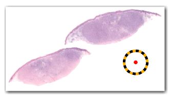
Figure 1.
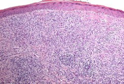
Figure 2.
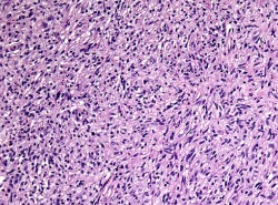
Figure 3.
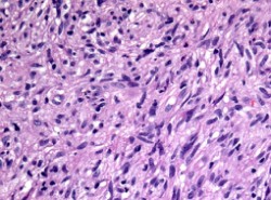
Figure 4.
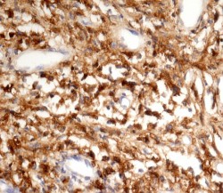
Figure 5.
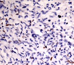
Figure 6.
What is the next best stain to perform?