Question 1:
A 34-year-old female presented with a slowly enlarging 3 x 4 cm ill-defined, cobbled, yellow plaque on her left lower abdomen. It was present since her early childhood. There was patchy hyperpigmentation within the plaque. A punch biopsy was performed. The main clinical differential diagnoses were connective tissue nevus, dermatofibrosarcoma protuberans versus other.
Click this image to view the virtual slide.
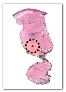
Figure 1. Punch biopsy skin 2x
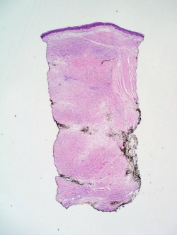
Figure 2. Punch biopsy skin 10x
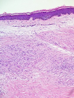
Figure 3. Punch biopsy skin 20x
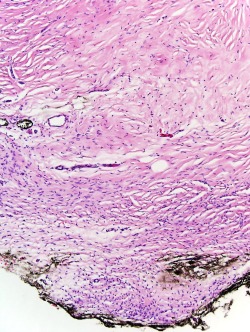
Figure 4. Punch biopsy skin 60x
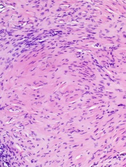
Figure 5. Punch biopsy skin Factor XIIIa
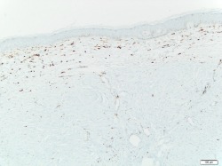
Figure 6. Punch biopsy skin Factor CD34
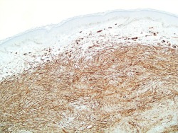
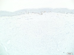
Figure 7. Punch biopsy skin Factor S100
What is the most likely diagnosis?