Question 1:
A two-month-old infant boy presented with a right frontal scalp mass fixed in position to the skull. He was the product of a full-term gestation and delivered via cesarean section due to fetal distress. Skull X-ray showed a 2 cm lytic lesion with irregular sclerotic margins in the right parietal bone. This was followed by a complete skeletal survey with no other identifiable lesions and a head CT with contrast that highlighted the lytic lesion, but revealed no intracranial breakthrough lesions or abnormal contrast enhancement (Figure 1). The differential diagnosis included an eosinophilic granuloma (Langerhans cell histiocytosis) and a resolving calcified cephalohematoma. The lesion was asymptomatic to the child causing no neurologic sequelae, but had increased in size. At age 6 months, the mother opted for complete excision of the mass. The histopathologic findings are presented in the figures below (Figures 2-6).
Click this image to view the zoomable slide.
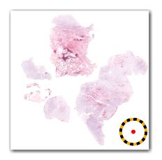
Figure 1.
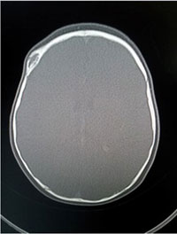
Figure 2.
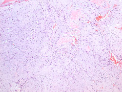
Figure 3.
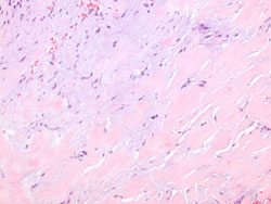
Figure 4.
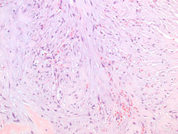
Figure 5.
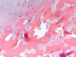
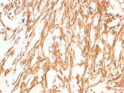
Figure 6. Stain: Smooth muscle actin
Which of the following is the best diagnosis?