Question 1:
An otherwise healthy 28-year-old male presented with a 1-year history of a painful lesion on his left abdomen. He had been applying a topical benzoyl peroxide-clindamycin gel to the area for 6 months without improvement. Visual inspection revealed a 4mm well-defined, pink papule (Figure 1). Palpation further revealed a tender indurated plaque as well as 2 additional tender papules that were not visible on exam. An 8mm punch biopsy was performed on the visible papule; the histopathology is demonstrated in Figures 2-5.
Click this image to view the virtual slide.
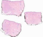
Figure 1 - Well circumscribed pink papule on the left abdomen

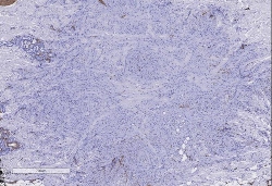
Figure 2 - Hematoxylin and eosin 4x
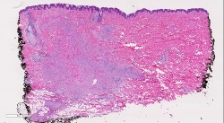
Figure 3 - Hematoxylin and eosin 10x
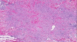
Figure 4 - Hematoxylin and eosin 40x
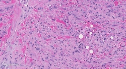
Figure 5 - FOSB Stain 40x
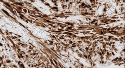
Figure 6 - ERG Stain 20x
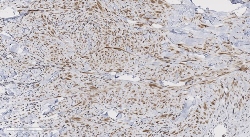
Figure 7 - CD34 Stin 10x
Which of the following is the most likely diagnosis?