Question 1:
A 61-year-old female presents with a long standing history of a wart-like plaque on the posterior aspect of her left calf. She states that the lesion had been present since birth and documented by her dermatologist as congenital. It was biopsied 10 years ago, and diagnosed as a hemangioma by an outside laboratory. Following biopsy, the lesion eventually recurred at the same site and continued to grow, although specific details with respect to the time frame were not documented. The lesion occasionally bled, but was not ulcerated. There were no other visible lesions in the surrounding region.
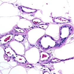
On examination the patient had a 1.3 cm purple, dome–shaped, crusted nodule. An excision was performed, the histopathology of which is pictured in Figures 1-4.
Click this image to view the virtual slide
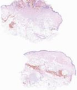
Figure 1
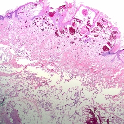
Figure 2

Figure 3
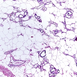
Figure 4
Which of the following is the best diagnosis?