Question 1:
A 7-month-old male presented with a right arm mass that had been growing for the past 4 months. Clinical differential diagnoses included lipoma, hemangioma, or rhabdomyosarcoma and the imaging studies including MRI, ultrasound, and doppler flows suggested the lesion to be either a hemangioma or rhabdomyosarcoma. A biopsy of the lesion revealed the following histopathologic changes:
Click the picture below to view the virtual slide.
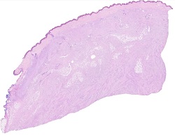
Figure 1.
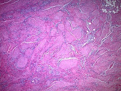
Figure 2.
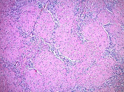
Figure 3.
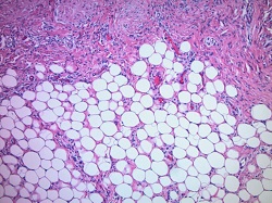
Figure 4.
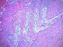
Which of the following is the best diagnosis?