Question 1:
A 47-year-old male presents to his dermatologist with a 1.1 x 1.0 cm flesh-colored papule on the left index finger near the nail bed. The lesion has been present for two years, has been gradually increasing in size, and does not cause pain. It is firm and solid to palpation. An excisional biopsy is performed.
Click the image below to view the virtual slide.
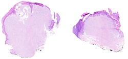
Figure 1.
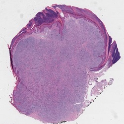
Figure 2.
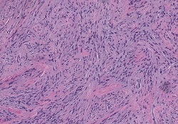
Figure 3.
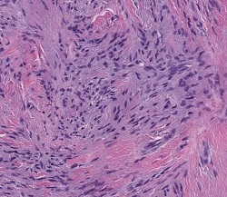
Figure 4. EMA
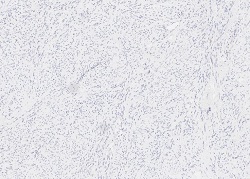
Figure 5a. CD34
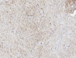
Figure 5b. CD34
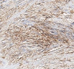
Figure 6. Pankeratin
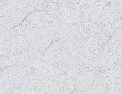
Figure 7. S100

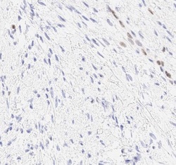
Figure 8. Rb1
Which of the following is the best diagnosis?