Question 1:
A 36-year-old women presents with a 3-month history of new skin findings affecting her left hip. Clinical exam reveals a painless, well-circumscribed, atrophic-appearing plaque with a faint violaceous hue, which was most prominent at the periphery of the lesion (Figure 1). She denies any significant medical history or recent injury. A punch biopsy of the area showed the following histopathology (Figure 2 - 6). An additional punch biopsy of nearby normal skin was also performed (Figure 7 - 8).
Figure 1.
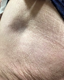
Figure 2.
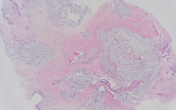
Figure 3.
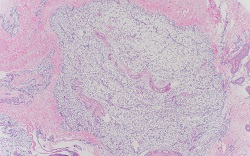
Figure 4.
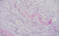
Figure 5.
.jpg)
Figure 6.
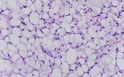
Figure 7.
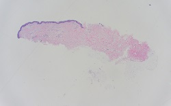
Figure 8.
.jpg)
What is the most likely diagnosis?