Question 1:
A 67-year-old man with history of immunoglobulin M (IgM) monoclonal gammopathy presented with a 2-centimeter, asymptomatic erythematous plaque with papules, prominent telangiectasias and a faint orange hue on the left posterior shoulder.
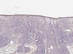
The lesion was biopsied, H&E and immunohistochemical (IHC) stains for CD3, CD20, CD138, PAX5, kappa, and lambda light chains were performed. The images are shown below. A CD5 stain, performed on a prior biopsy, highlighted only T lymphocytes.
Click this image to view the virtual slide
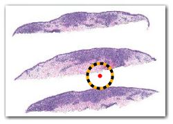
Figure 1. Clinical Lesion

Figure 2. H&E, 1x
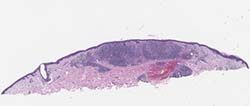
Figure 3. H&E, 12x
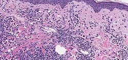
Figure 4. H&E, 40x
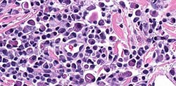
Figure 5. CD3, 20x
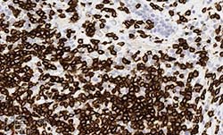
Figure 6. CD20, 20x
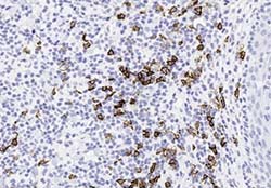
Figure 7. CD138, 20x
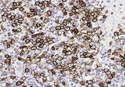
Figure 8. PAX5, 20x
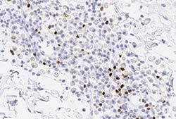
Figure 9. Kappa, 20x
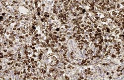
Figure 10. Lambda, 20x
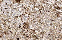
Figure 11. Congo red, 4x
What is the most likely diagnosis?